Marja Pitkänen’s doctoral thesis at Aalto University, defended on June 12, 2020, presents a groundbreaking exploration in the field of neuroscience and biomedical engineering. Titled “Ex vivo and corneal electroretinogram in temperature determination and functional characterization of the retina,” the dissertation unveils a novel application of electroretinography (ERG) to monitor retinal temperature changes. This technique, leveraging the temperature-sensitive nature of ERG responses, offers potential advancements in the safety and efficacy of retinal disease treatments by ensuring therapeutic temperatures are maintained without causing cellular damage.
https://urn.fi/URN:ISBN:978-952-60-3924-4
Abstract:
The first steps in vision take place in the layered tissue of the retina converting changes in incoming photon flux into neural signals and processing the signals in its neural network. Electroretinography (ERG) provides an experimental approach for studying retinal functions. ERG records light-induced changes in the extracellular field potentials of the retinal tissue. In the corneal or in vivo method, ERG signal is recorded non-invasively from the surface of the eye. By the ex vivo method, corresponding signal can be recorded from an isolated retina. The present work consists of studies applying the combination of these complementary experimental tools. The goal is to characterize retinal functions by ERG at varying temperatures and extracellular conditions and to investigate the feasibility of ERG for retinal temperature estimation.
The preservation of rod function in the isolated retinal tissue was assessed by investigating ERG responses ex vivo and comparing them to corresponding in vivo recordings with the retina in its physiological environment. According to the obtained results, rod function is well conserved in the isolated retina. The activation phase was similar and only minor reduction in rod sensitivity and decrease in photoresponse kinetics were observed ex vivo compared to in vivo. Thus, ex vivo ERG provides an applicable and reliable method for studying rod photoreceptor function and phototransduction.
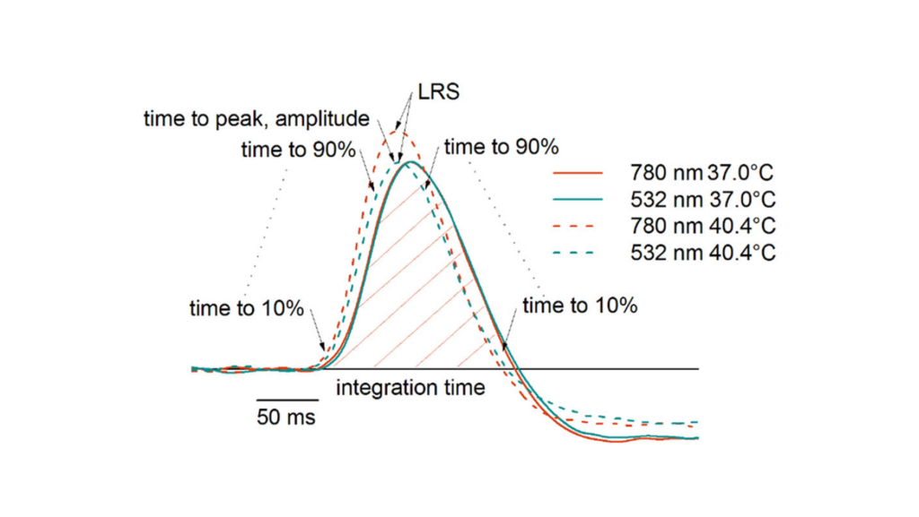
Investigating the temperature-induced changes in dark-adapted ERG flash responses constituted the most prominent part of this thesis. The study was motivated by the need of a retinal temperature monitoring method for controlling fundus heating treatments of retinal diseases. The changes in ERG responses due to thermal alterations near mammalian body temperature were quantified first in the ex vivo setup enabling fast and accurate temperature adjustments. Thereafter, the temperature induced changes in ERG parameters were adderessed by corneal ERG. Temperature estimation models were developed to demonstrate a proof of concept for the determination of retinal temperature based on ERG photoresponses. The RMS error level obtained with the method for corneal ERG was < 0.7 °C, applying recordings performed in a time interval of ~20 seconds.
The combination of ex vivo and corneal ERG was further applied for characterizing the retinal functions of a novel mouse model lacking P4H-TM enzyme. The corneal recordings indicated a defect in the cone-pathway of the retinas of these mice. However, as the function of the inactivated gene is related to the oxygenation level of tissues, and general anesthesia is known to cause abnormalities in the oxygen regulation in the body, ERG recordings were conducted also ex vivo in controlled environment with stable oxygen levels. Ex vivo ERG verified that the observed alterations in the photoresponses were caused by cone-mediated deficiency, and not by a mechanism related to the anesthetized state.