Researchers at Aalto University, Karolinska Institutet / St. Erik Eye Hospital, and the University of Helsinki demonstrate that real-time electroretinography (ERG)-based thermal dosimetry can activate cytoprotective pathways without retinal damage in a preclinical pig model.
“Non-damaging laser treatment with electroretinography-based thermal dosimetry activates hormetic heat response in pig retinal pigment epithelium”
https://doi.org/10.1038/s41467-025-64095-6
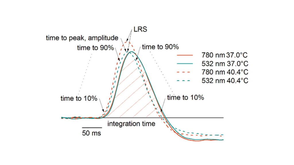
Espoo, Finland – October 29, 2025 – A study published today in Nature Communications reports that retinal laser exposures can be thermally dosed in real time using ERG, enabling non-damaging, temperature-controlled treatment in vivo (pig). The work, conducted by researchers at Aalto University, Karolinska Institutet / St. Erik Eye Hospital, and the University of Helsinki, shows that maintaining retinal temperature near 44 °C during 60-second near-infrared laser exposures increased HSP70/HSP90 and autophagy markers in the RPE/choroid while avoiding oxidative stress, apoptosis, and visible lesions. Visible lesions occurred above 48 °C, and temperature-dosing precision at the lesion threshold was approximately 0.6 °C.
Key points (preclinical):
Real-time thermal dosing: ERG-based dosimetry individualized laser power with ~0.6 °C precision at the lesion threshold temperature.
Therapeutic window: Targeting ~44 °C activated heat-shock and autophagy responses without structural damage.
Safety boundary: Lesions were observed above 48 °C, delineating a practical non-damaging vs damaging range.
The findings provide a quantitative framework for dosing non-damaging retinal laser therapies and motivate clinical evaluation of the presented novel temperature-controlled approach.
Abstract:
Enhancing protein homeostasis and antioxidant defense mechanisms in the retinal pigment epithelium (RPE) holds significant promise as a treatment option for various retinal diseases, including age-related macular degeneration. However, patient responses to laser-induced RPE hyperthermia varies substantially. To address this, we introduce a focal electroretinography (fERG)-based method for retinal temperature monitoring during laser exposure. Applying the method to anaesthetized male pigs in vivo, we study the biological effects of controlled retinal hyperthermia. Our findings reveal that temperature elevation to 44 °C with 60-second laser exposure triggers heat shock protein production and autophagy activation in RPE/choroid while avoiding oxidative stress, apoptosis, and structural damage. Importantly, our results demonstrate that visible lesions occur at temperatures above 48 °C, and that the temperature determination precision was 0.6 °C. These outcomes highlight that fERG-controlled retinal laser treatment enables reliable and safe activation of cytoprotective mechanisms in the RPE, providing a promising new therapeutic approach.